 Dentist Channel Online
Horizontal Bone Loss
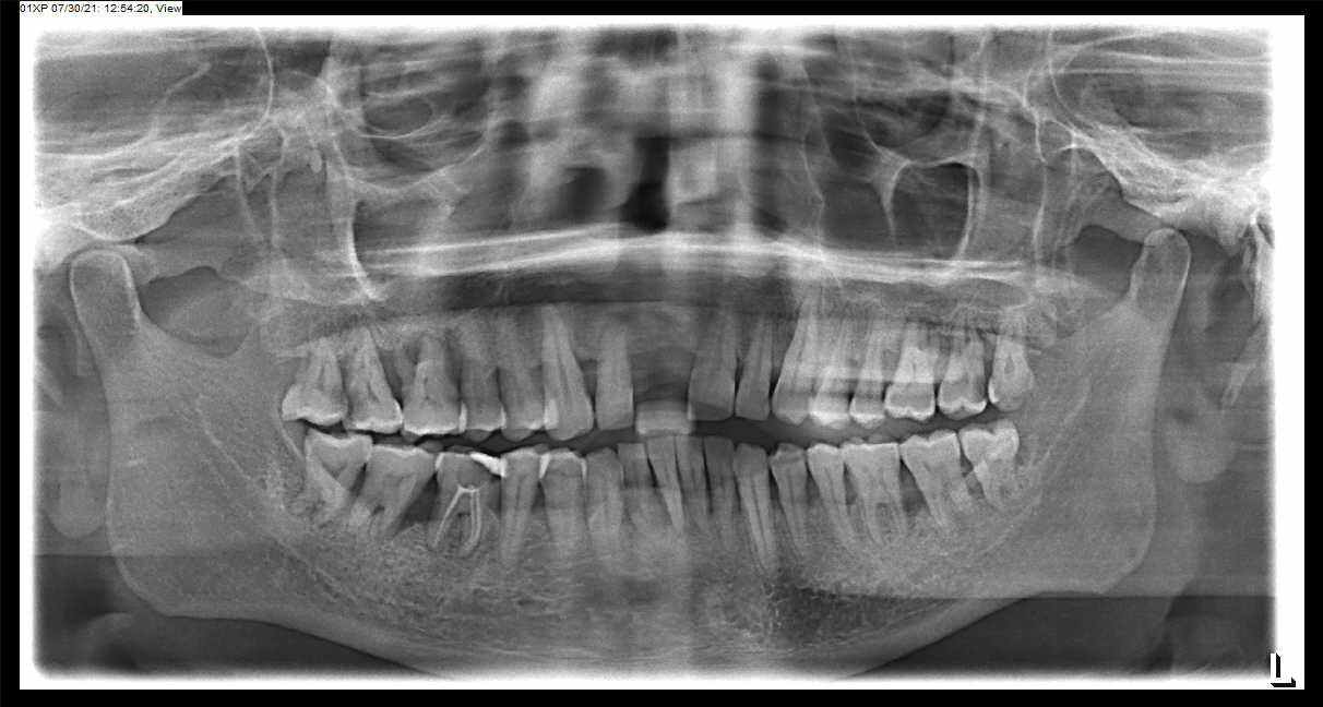
Radiographic appearance of loss in height of the alveolar bone where the crest is still horizontal but is positioned apically more than a couple of millimeters from the CEJs. Horizontal bone loss may be mild, moderate, or severe, depending on its extent.
Mild bone loss may be defined as approximately a 1- to 2-mm loss of the supporting bone. Moderate loss is greater than 2 mm up to loss of half the supporting bone height. Severe loss is beyond this point.
Vertical Bone Defects
The term vertical or angular osseous defect describes a bony lesion that is localized to a single tooth, although an individual may have multiple vertical osseous defects. These defects develop when bone loss progresses down the root of the tooth, resulting in deepening of the clinical periodontal pocket.
The radiographic presentation is a vertical deformity within the alveolus that extends apically along the root of the affected tooth from the alveolar crest. The outline of the remaining alveolar bone typically displays an oblique angulation to an imaginary line connecting the CEJ of the affected tooth to the neighboring tooth.
The vertical defect is described as three walled (surrounded by three bony walls) when both buccal and lingual cortical plates remain. It is described as two walled when one of these plates has been resorbed. One walled when both plates have been lost. The distinctions among these groups are important in designing the treatment plan.
Often vertical defects are difficult or impossible to recognize on a radiograph because one or both of the cortical bony plates remain superimposed over the defect.
Interdental Craters
Two-walled, trough like depression that forms in the crest of the interdental bone between adjacent teeth. Radiographically, bandlike or irregular region of bone with less density at the crest, immediately adjacent to the more dense normal bone apical to the base of the crater.
Buccal or Lingual Cortical Plate Loss
Loss of a cortical plate may occur alone or with another type of bone loss such as horizontal bone loss. Increase in the radiolucency of the root of the tooth near the alveolar crest.
Osseous Deformities in the Furcations of Multirooted Teeth
Very early furcation involvement of a mandibular molar characterized by slight widening of the periodontal ligament space in the furcation region.
The bony defect may also involve only the buccal or lingual cortical plate and extend under the roof of the furcation. In such a case, if the defect does not extend through to the other cortical plate, it appears more irregular and radiolucent than does the adjacent normal bone.
In the mandible, the external oblique ridge may mask furcation involvement of the third molars. Convergent roots may also obscure furcation defects in maxillary and mandibular second and third molars. The image of furcation involvement is not as sharply defined around maxillary molars as around mandibular molars because the palatal root is superimposed on the defect.
Radiographs should be taken at different angles to reduce the risk of missing furcation involvement.
Article by Dr. Siri P. B.

Hailey - 9 months ago