So is it true, that a simple X-ray can cause cancer…?
While high doses of X-rays and other types of high-energy electromagnetic radiation are known as highly cancerous, most studies haven’t found a connection between exposure to low levels of radiation
Most of the evidence linking high levels of radiation exposure to cancer comes from studies of certain groups of people, such as people who survived the atom bomb blasts in Hiroshima and Nagasaki in Japan during World War 2, survivors of the Chernobyl nuclear accident in Russia (1986), and people like uranium miners and nuclear power plant workers whose jobs expose them to high amounts of radiation.
As the risk of cancer rises with the level and duration of exposure, such cases cannot be taken into account when talking about medical X-rays.
Children have been known to be much more prone to radiation and to develop long-term health problems as a result of radiation therapy and therefore are prescribed lower dosages of radiation-based treatments than adults.
Although many epidemiological studies have reported on a connection between exposure to dental X-rays and risk of brain tumor, it is still controversial, as some studies have shown that this might not be true. However, studies have shown that there is no clear evidence on the existence of a significant connection between exposure to dental X-rays and the risk of developing a Brain tumor
In many cases, Thyroid cancer is amongst the most common cancers worldwide, and exposure to dental radiation is likely to contribute to its occurrence due to the location of the thyroid gland, as the thyroid is one of the most susceptible parts of the body repeated exposure to dental X-rays may result in various health problems like head and neck tumors and various systemic problems.
Summary
Thus, we summarize that all of those theories are partially true as it is true that radiation can indeed cause cancer, it is unclear however as to what amount of radiation would be enough to cause cancer, and it is also true that the patient’s resistance to radiation plays a key role here, but if we look at the number of patients who receive a Dental X-ray every year statistics show us another result altogether.
Therefore, the conclusion is that X-rays are safe as long as the correct tests and procedures are run on the patients before taking an X-ray, giving the doctor information about the patient’s immunity to diseases, or possibly in the near future their resistance to radiation, this will allow the doctor to avoid radiation therapy for those patients who have less tolerance towards radiation and diseases
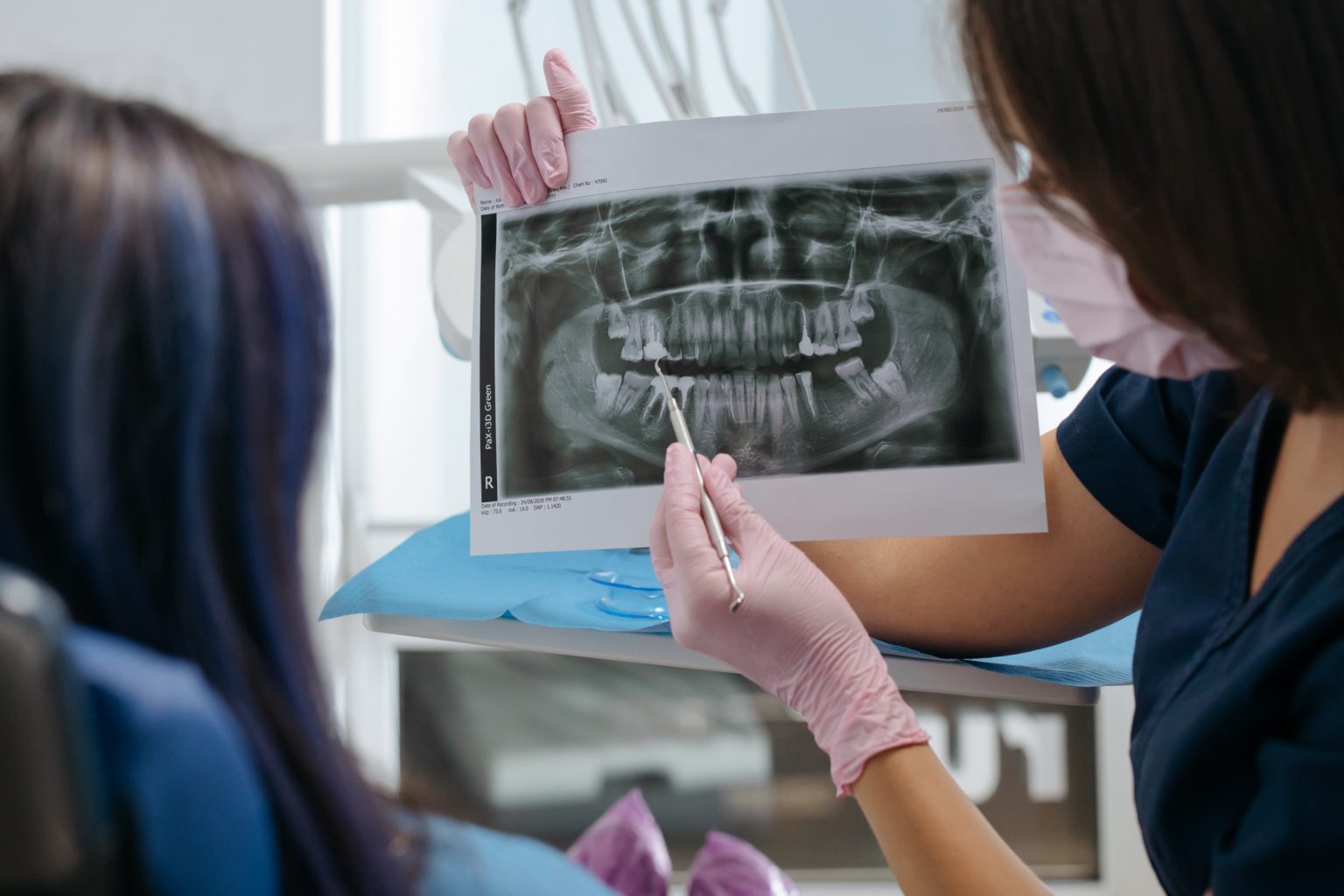
Doctor Diagnosing
How does an x-ray machine work…?
An x-ray machine produces x-rays and directs them toward an object. The x-rays then pass through some parts of the object but are blocked by other parts. For example, x-rays directed at a body pass through the skin and soft tissues, such as muscle. A special film placed behind the body captures the image made by the x-rays.
Dentists use x-rays to check the health of the patient’s bones, teeth, fillings, crowns, or implants they may have. But, not all dental x-rays are the same.
Types of Dental X-rays
Dentists use various types of x-rays machines, depending on what they’re trying to find, here are the five most common dental x-rays machines used for dental X-rays
Bitewing X-rays
A bitewing x-ray is used to look at certain areas of your mouth, a dentist may request one or more bitewing x-rays during a check-up.
Each bitewing captures the visible parts of your upper and lower teeth as well as half of their roots and supporting bone, and such x-rays help dentists to detect decay, especially between teeth.
This helps dentists detect changes in the structure of your jawbone caused by gum disease.
In most cases, the patient will be asked to bite down on a piece of plastic that holds the x-ray film against their upper and lower teeth, if the dentist uses digital x-rays, the patient will be asked to bite down on a little box that’s wrapped in plastic.
Periapical X-rays
A periapical x-ray captures the whole tooth. It shows everything from the crown to the root, each periapical x-ray shows a small section of your upper or lower teeth, such x-rays are often used to detect any unusual changes in the root and surrounding bone structures.
The film will be placed near to the patient’s mouth using a metal rod with a ring attached to it, the patient will need to bite firmly onto the device to keep it in place and in order to provide a clear x-ray image.
Full Mouth X-rays Survey
A full mouth x-ray survey is composed of a series of individual images, including a combination of bitewing and periapical.
Dentists use these initial images as a baseline on the health of your mouth, although full mouth x-ray surveys are usually only taken when your dentist suspects you have a jaw cyst or tumor, they’re also used for dental work such as root canals, extractions, and gum disease treatment.
The patient will be asked to take bitewing and periapical x-rays simultaneously.
Cone Beam Computed Tomography (CBCT)
CBCT is a radiographic imaging method that allows to accurately create 3D imaging of hard tissue structures and is capable of providing images of higher diagnostic quality, with short scanning times.
The CTCB machine has a motorized arm that rotates 360-degrees around the patients head while capturing multiple images from different angles that are then reconstructed to create a single 3D image.
Panoramic X-rays or (PR)
Panoramic x-rays, are used to take images of your entire mouth area, that show the position of exiting, emerging, and impacted teeth, all in one image, panoramic x-rays also called as panoramic radiography is a form of focal plane tomography, which shows a 2D view of a half circle from one ear to the other displaying the entire structure of the jaw like a horseshoe.
The patient will be asked to bite down on a gag or a bite locker to keep their teeth aligned in order to obtain a clear image. Then, a rotating arm on the machine makes a semi-circle around your head to record your mouth.
Occlusal X-rays
Occlusal x-rays help track the development and placement of a section or entire arch of teeth in the upper or lower jaw, they’re mostly used by pediatric dentists to find a child’s teeth that have not yet broken through the gums.
The process is very similar to a bitewing x-ray.
What type of X-ray machines are better for Dental use…?
Generally, x-ray machines that use DC current are better than the AC x-ray machines.
AC X-ray Machines:
An AC x-ray machine is one of the cheapest ways to produce x-rays, which uses the alternating current from the electricity provider to power the x-ray tube head.
The reason for using such a current is to produce x-rays that turn on and off sixty times each second as for the x-rays that are produced, each begins too weak to use and grow to proper strength before they diminish again to being too weak.
Pros
This is one of the cheapest ways to produce X-rays.
Cons
This type of design of AC X-rays requires filters of aluminum to eliminate the rays that are too weak to use.
DC X-ray Machines:
A DC x-ray machine uses a direct electric current to power the x-ray tube head. These constant potentials of DC x-ray machines, neither change the direction nor the intensity making it a steady supply of power. This facilitates the production of smooth and consistent x-rays.
Pros
Consistent x-ray images.
Reduced risk Exposure to Radiation.
Constant Voltage values.
Cons
This type of x-ray is slightly expensive.


Hailey - 1 year ago