
 Dentist Channel Online
The role of a Laser in Implant dentistry
Dr. Kirpa Johar
B.D.S, Masters Certification in Laser Dentistry (Vienna)
Director, Haridaan Clinic and Academy, Bengaluru, India
Implant dentistry represents a viable alternative for many patients in need of a removable prosthesis. Dental lasers aid in improving the presurgical, surgical, postsurgical and prosthetic phases of implant dentistry. The biological seal must be effective enough to prevent the ingress of bacterial plaque, toxins, oral debris and other deleterious substances. If a biological seal is created from the beginning of implant uncovering using laser technology, the attached gingiva would heal directly around the implant, forming an epithelial cuff.
PREPARATION OF THE SURGICALSITE
It is the first step of implant surgery. Lasers present an excellent solution to the problem of surgical site contamination. All lasers are bactericidal. The bactericidal effect is profound and almost instantaneous, and an implant site can be sterilized. Before osteotomy development, the soft tissue can be sterilized much more effectively with a laser than with rinsing or swabbing. Further, if saliva were to contaminate the surgical site during the surgical procedure, it would be simple to release the area and re-establish sterility so that the procedure can continue with the greatest chance for success.
DECONTAMINATION AND IMPLANT PLACEMENT
A CO2 laser has a distinct advantage over contact lasers. Because CO2 lasers are non-contact, it is a simple procedure to place a wide-angle handpiece on the CO2 laser and use it out of focus, which would increase the spot size on the tissue even more. The clinician should gently curette the bone to re-establish bleeding and maximize the healing potential of the implant or graft site. The laser energy must be delivered to all the bony surfaces within the extraction site. If a severely dilacerated root causes an inaccessible line of sight for the access of the laser beam, the clinician should allow the body’s natural defense mechanism to heal the site over weeks and make it safe for re-entry to perform the implant or grafting procedure.
SOFT TISSUE OSSTEOTOMY
The next objective in laser implant surgery is preparation of the osteotomy through soft tissues first and then the hard tissues. The clinician must first decide on the desired pattern of entry through the soft tissue. In some cases, a minimal entry referred to as “punch procedure” is the goal. The soft tissue is removed in a 3-4mm diameter down to the crest of bone. This soft tissue may be 1-2 mm or 3-4mm thick depending on the location and the biotype. If the tissue is as thin as l-2mm, any wavelength is acceptable, but if its thicker, using a diode or an Nd: YAG may take more time versus seconds for erbium and CO2 lasers. The unobstructed vision, excellent hemostasis and efficiency of cutting through all tissue biotypes and thickness make the CO2 laser most suitable for these procedures.
DIODE LASERS FOR MUCOSITIS PERI-IMPLANTITIS
Mucositis is a soft tissue infection around the abutment-crown-implant complex, typically at the cervical third of the implant. Peri-implantitis is an infection around the body or apex of the implant that leads to loss of bone. Both these conditions are caused by an inflammatory reaction to anaerobic plaque bacteria associated with a biofilm. This results in swelling and inflammation of the soft tissues and bone loss surrounding the implant. Before any laser surgery is attempted, the initial surgery and healing process should be evaluated. A rigidly placed implant with no crestal bone loss and adequate zones of attached gingiva should be present. Soft tissue thickness of l-3mm with no tenderness or discomfort under vertical or lateral forces is needed. Once the implant is uncovered with the laser under minimal anesthesia, the rigidity can be verified.
The soft tissue greater than 3mm thick should be reduced with the laser to create an ideal pocket depth around the implant. If bone defects are encountered or the width of attached tissue is less than 3mm, full surgical reflection is recommended.
Soft Tissue Diode Laser as an adjunct to Implant Dentistry
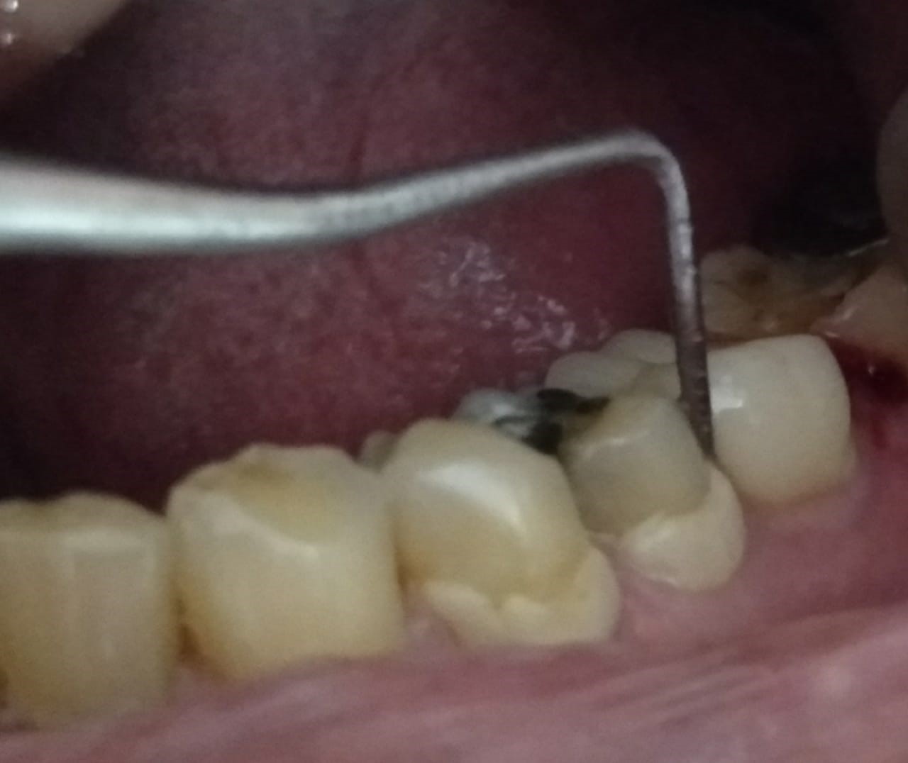
Case Study: A 65-year-old female patient came to the dental operatory with gingival inflammation and bleeding around tooth no 36.
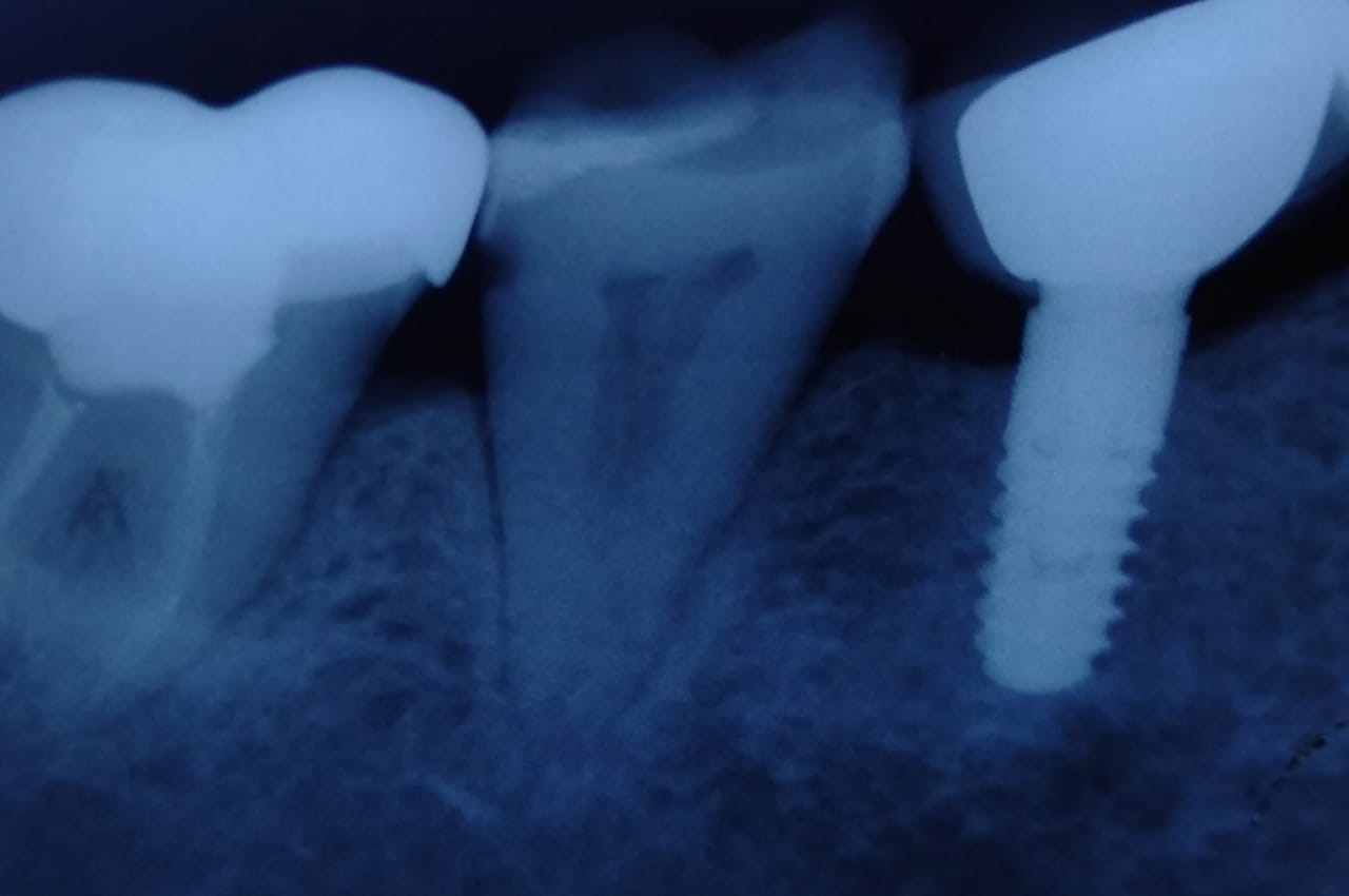
IOPA radiograph of the same
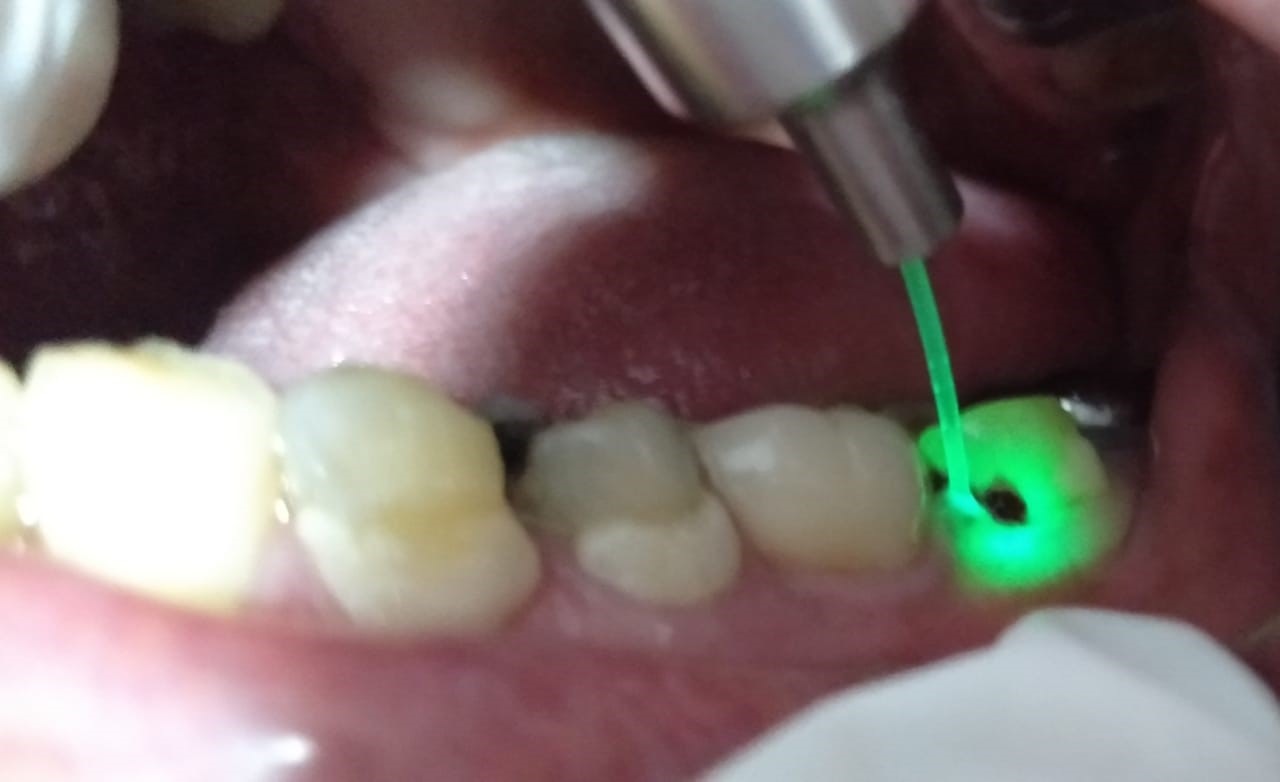
Laser assisted peri-implantitis treatment around tooth no 36.
ADVANTAGES OF USING LASERS
Using laser energy to make any incision has several benefits:
A sterile cut is less likely to become infected.
Lasers incise tissue without creating the cascade of events that leads to swelling and inflammation. Healing is enhanced because of reduced inflammation and postoperative pain.
For erbium lasers to replace the current Implant drills, precision in depth and diameter of cut is necessary.
In another area, low level laser application is thought to improve wound healing, with evidence of accumulated collagen fibrils, accelerated cell reproduction and increased prostaglandin levels.
CONCLUSION
Lasers have significant benefits in modern clinical dentistry, especially in implant dentistry.
Diode, CO2 and erbium lasers have the potential to improve the clinician’s ability to deliver the highest quality of care, while providing a more comfortable experience for the patient with fewer postoperative problems.


Hailey - 9 months ago