• Dentist Channel Online Dentist Channel Online
  • 8 September, 2021

Structure of Tooth - Basic Anatomy that all Must Know

Structure of Tooth - What are the different layers ?

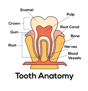
The outermost layer in the structure of tooth is made up of enamel. It it's the hardest and most mineralised substance of the body. It's the visible part of the tooth. This layer of tooth is most susceptible to dental caries, attrition / non-carious cervical lesions and fracture. This layer can never regenerate. Repair by itself is impossible but possible only when the dentist intervenes. The main inorganic content of enamel is hydroxyapatite crystals. Inorganic content constitutes up to 96 percent and 4 percent of organic content.  

The structure underneath the enamel of the tooth is dentine. The organic content is more than enamel. Major part i.e. up to 60 percent of dentin is made up of inorganic content and rest by organic. This layer can generate reparative dentin. It's yellowish in colour. It's sensitive due to presence of dentinal tubules traversing from the pulp to the dentine. These dentinal tubules carry dentinal fluid. The caries process is more damaging here than enamel 


The innermost layer of the crown structure is pulp. This layer consists of nerves and blood vessels. Any damage to the enamel followed by to dentin if it reaches pulp, the tooth starts aching and further there is chances of loosing its vitality. The pulp and the dentine share a close relationship. The cells in these both layers are associated and function as a unit. Cell processes from pulp extend to dentin. The pulp can be called as connective tissue containing cells, ground substances, fibres, interstitial fluid, cellular components etc. The pulp receives blood and nerve supply through the apical foramen of the tooth. Apical foramen forms the apex of the tooth. It consists of major and a minor opening / diameter. Apical region of the root carries lot of significance 

The supporting structure surrounding the tooth are periodontal ligaments, alveolar bone. Tooth root is covered by cementum. The periodontal ligament consists of various fibres which anchors the tooth to the underlying and surrounding alveolar bone. Alveolar bone forms the socket for the tooth. The cementum in the root protects the dentin and the pulp. Any damage to the surrounding tissues of the teeth requires intervention of periodontist who is specialised in taking care of health and damage to these structures 

Take home message 

It's important to take care of tooth as well as surrounding structures since they work as an unit. Tooth is that tissue if once lost cannot be regained. Only prosthesis can be given as substitute but can never function as effectively as the natural tooth. 

Article by Dr. Siri P.B.


Categories:
Tags :
Comments :
Hailey Avatar
Hailey - 8 months ago
My name is Hailey Garcia and I am from New Jersey. My herpes virus turned to war after 2 years of living with it. I have tried different medical procedures to cure my herpes but to no avail. Most people think herpes is only a minor skin irritation of which herpes has long term effects on health and passes through the bloodstream and can be easily contracted through sexual intercourse. I knew I had herpes from the first day I started feeling itchy in my pubic area and the pain was very unbearable. I couldn't stand it anymore. After 2 years of trying other means to get rid of it, I had to contact Doctor Odunga to help me with a permanent cure. I saw his email and whats-app number from a testimony I read online from a lady who was also helped by him in curing infertility problems, I had faith and contacted him. He assured me of his work and I ordered his herbal medicine. Within 5 days, I didn't feel any pain anymore and within 2 weeks, my skin was all cleared and smooth. I am very grateful to you sir and I write this testimony as others have done to bring those having faith to you sir. If you have herpes or other similar disease and you want it cured, kindly contact Doctor Odunga, Whats-App (wa.me/+2348167159012) OR Email [email protected]

Authentication required

You must log in to post a comment.

Log in

Upcoming webinars:

  • 18 December,2025
Shoot, Document, Deliver: The Power of Dental Photography

Join our webinar ‘Shoot, Document, Deliver: The Power of Dental Photography’ to master equipment, techniques, and applications for impactful clinical images.

  • 21 December,2025
Minor Oral Surgery, Major Challenges : Clinician’s Guide

Gain practical insights into managing common challenges and complications in minor oral surgery for improved clinical outcomes.

  • 25 December,2025
From failing dentition to fixed function: Advanced full-arch implant restorations with a patient-centered approach

Discover advanced full-arch implant restorations transforming failing dentition with a patient-centered approach.

  • 28 December,2025
In the Loop : Decoding the Matrix Systems

Simplifying the matricing challenges from Flat to Fabulous

  • 1 January,2026
Management of Complications Post Transalveolar Extractions.

This Webinar will cover Management of Various Complications Occurring after Surgical Extractions.

  • 2 January,2026
Basics of Prosthetic Options in Dental Implants

Choosing the right prosthesis is the key to long-term success in implant dentistry.

  • 16 January,2026
Abutment Selection In Implantology- Which, When and Why?

This webinar will briefly discuss various abutment options and their selection process to enhance implant function and longevity.

  • 18 January,2026
Clinical Implications of Proximity of Nerve with Mandibular Third Molars

A Webinar on Evaluating Nerve Proximity Risks during Mandibular Third Molar Surgery for safer Clinical Outcomes.