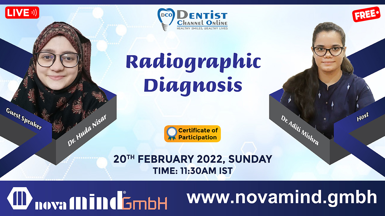
Radiographic Diagnosis

OBJECTIVE :  
Being able to detect dental caries on radiographs is an essential skill needed for providing comprehensive dental treatment. Certain types of dental caries are difficult to visualize intraorally, and therefore, the diagnosis need to be made based solely on the radiographs. This module will introduce students to a larger number of radiographic examples of caries than typically covered in lecture. 

Dental radiographs are critical for the complete assessment and treatment of dental diseases. Dental radiography is commonly used to evaluate congenital dental defects, periodontal disease, orthodontic manipulations, oral tumors, endodontic treatments, oral trauma, and any situation where an abnormality is suspected. Although standard radiographic equipment and film can be used to produce dental radiographs, dental X-ray equipment and film provide superior quality images and greater convenience of animal patient positioning. An understanding of normal dental radiographic anatomy is important when interpreting dental radiographs. Stage III periodontitis is the earliest stage of periodontal disease at which radiographic abnormalities become apparent. Bone loss associated with periodontal disease can be classified as either horizontal or vertical. Periapical radiolucencies can represent granulomas, cysts, or abscesses, whereas periapical radiodensities may represent sclerotic bone or condensing osteitis. Lytic lesions of the bone of the jaw often represent oral neoplasms. Neoplasms also can displace or disrupt teeth in the dental arch. Resorptive lesions can be external or internal and appear as radiolucent areas involving the external surface of the root or the pulp cavity, respectively.  
 

WHAT WILL YOU LEARN IN THIS SESSION ?  

  1. Introduction
  2. Basics of Radiology
  3. Tooth associated pathology
  4. Bone associated pathology
  5. Difference between commonly misinterpreted lesions
  6. Conclusion
  7. Live Q & A  

DATE : SUNDAY, 20th FEBRUARY, 2022 

TIME : 11.30 AM IST  


Watch Recorded Video :

Speaker(s)

Categories:
Tags :
Comments :
There are no comments yet.
Authentication required

You must log in to post a comment.

Log in
Free Webinar
  Start Date

20 February, 2022 11:30 am

  End Date

20 February, 2022 12:30 pm

  Location

India

  Platform

Zoom

-

Advertisement

Related Post:

  • 14 February,2025
Apec Dhaka 2025

The 23rd Scientific Congress of Asian Pacific Endodontic Confederation

  • 4 February,2025
AEEDC 2025 | A Global Platform for the Dental World |29th Edition

AEEDC Dubai continues its tradition of bringing together the suppliers and buyers to conduct serious business and learn new techniques.

  • 24 November,2024
Integrating Periodontics into a Thriving Dental Practise: Strategies for Success

Integrating periodontics into a dental practice elevates patient care and significantly boosts practice growth

  • 17 November,2024
Single visit Endodontics- And Associated Myths Buster

A single sitting root canal can be challenging but doable

  • 16 November,2024
Integrating Laboratory Testing in Standard Dental Practice

Discover the essential laboratory investigations that ensure safe and successful general dental practice.

  • 15 November,2024
CAD/CAM Digital & Oral Facial Aesthetics 37th Int’l Dental Confex

On 15-16 November 2024, CAPP will host the CAD/CAM Digital & Oral Facial Aesthetics 37th Int’l Dental ConfEx

  • 10 November,2024
Mastering Contacts and Contours in Restorative Dentistry: A Comprehensive Guide

The presentation provides a comprehensive understanding of how to achieve optimal results in restorative situations.

  • 9 November,2024
TMDs Simplified

Join this webinar to explore temporomandibular joint disorders, diagnostic tools, conservative treatments, and practical solutions for better patient care.

  • 8 November,2024
The NPZ Course– Nasalis/ Pterygoid / Zygomatic : “All on 4 & All-on-X Technique

Master Zygomatic Implantology with our interactive 2-day hands-on model and live surgery workshop led by the Master of TILT.