• Admin
  • 23 April, 2022

Radiographic Assessment of Periodontium

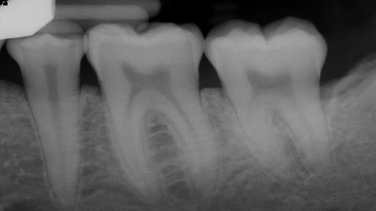
The main features to be looked out are for

  • Crown/ Root Ratio 
  • Open contacts 
  • Root length and morphology,  
  • Local irritating factors 
  • Amount of bone present 
  • Condition of the alveolar crests 
  • Bone loss in the furcation areas 
  • Width of the periodontal ligament space 

Normal Anatomy 

A thin layer of opaque cortical bone often covers the alveolar crest. The height of the crest lies at a level approximately 0.5 to 2.0 mm below the level of the CEJs of adjacent teeth. Between posterior teeth the alveolar crest also is parallel to a line connecting adjacent CEJs. Between anterior teeth the alveolar crest usually is pointed and may have a well-defined cortex. 

Between the anterior teeth, the normal alveolar crest is pointed and well corticated, coming to within 0.5 to 2.0 mm of the adjacent CEJs. The normal alveolar crest lies 0.5 to 2.0 mm below the adjacent CEJ and forms a sharp angle with the lamina dura of the adjacent tooth. 

The periodontal ligament (PDL) space is often slightly wider around the cervical portion of the tooth root, especially in adolescents with erupting teeth. In this situation, if the lamina dura still forms a sharp, well-defined angle with the alveolar crest, the condition is a variant of normal and is not an indication of disease. The thickness of alveolar crests varies and may be very thin coronally. This may appear radiographically as an increase in radiolucency toward the crest.  

The PDL varies in width from patient to patient, from tooth to tooth in the individual, and even from location to location around one tooth. Usually it is thinner in the middle of the root and slightly wider near the alveolar crest and root apex, suggesting that the fulcrum of physiologic movement is in the region where the PDL is thinnest. The shape of the tooth creates the appearance of a double PDL space. When the x-ray beam is directed so that two convexities of a root surface appear on a film, the double PDL space is seen. 

General Radiographic Features of Periodontal Disease 

  • Changes in the morphology of the supporting alveolar bone 
  • Loss of the interproximal crestal bone and bone overlapping the buccal or lingual aspects of the tooth roots
  • Changes to the internal density and trabecular pattern 
  • Changes to the internal aspect of the alveolar bone reflect either a reduction or an increase in bone structure or a mixture of both
  • Changes In Morphology Of The Alveolar Bone Appear as areas of localized erosion

 Article by Dr. Siri P. B.

Categories:
Tags :
Comments :
There are no comments yet.
Authentication required

You must log in to post a comment.

Log in

Upcoming webinars:

  • 23 November,2024
Navigating Dental Trauma: Clinical Insights and Treatment Protocols

The Webinar Explores Comprehensive, Evidence-Based Strategies for Diagnosing, Managing, and Preventing Traumatic Dental Injuries.

  • 24 November,2024
Integrating Periodontics into a Thriving Dental Practise: Strategies for Success

Integrating periodontics into a dental practice elevates patient care and significantly boosts practice growth

  • 24 November,2024
Dento-Facial Changes in Women after Menopause

Due to Hormonal changes after menopause, there are other factors which get altered in a female body.

  • 28 November,2024
Biological Bone Augmentation & The Semilunar technique:Only for Real Bone Builders

Biological Bone Augmentation with autogenous bone allows predictable implantologic results even in compromised bone with reduced bone volume.

  • 29 November,2024
Endodontics Demystified - Techniques that make sense

Endodontics Demystified” is a no-stress, high-impact webinar breaking down root canals with pro tips and real cases

  • 30 November,2024
TMDs Simplified

Join this webinar to explore temporomandibular joint disorders, diagnostic tools, conservative treatments, and practical solutions for better patient care.

  • 1 December,2024
Isolation and its significance in dentistry

Isolation in dentistry involves techniques to control moisture and contamination, crucial for infection control and enhancing treatment effectiveness.

  • 1 December,2024
Simvastatin in Periodontics

Current trends of application of simvastatin as LDD