 Dentist Channel Online
The term "smear layer" is used most often to describe the grinding left on dentin by cavity preperation. The term applies to any debris produced iatrogenically by the cutting, not only of dentin, but also of enamel, cementum and even dentin of the root canal.
Some phases of cosmetic dentistry demands that depending on what dentin bonding agent is used, the smear layer be retained, other material dictates it's removal.
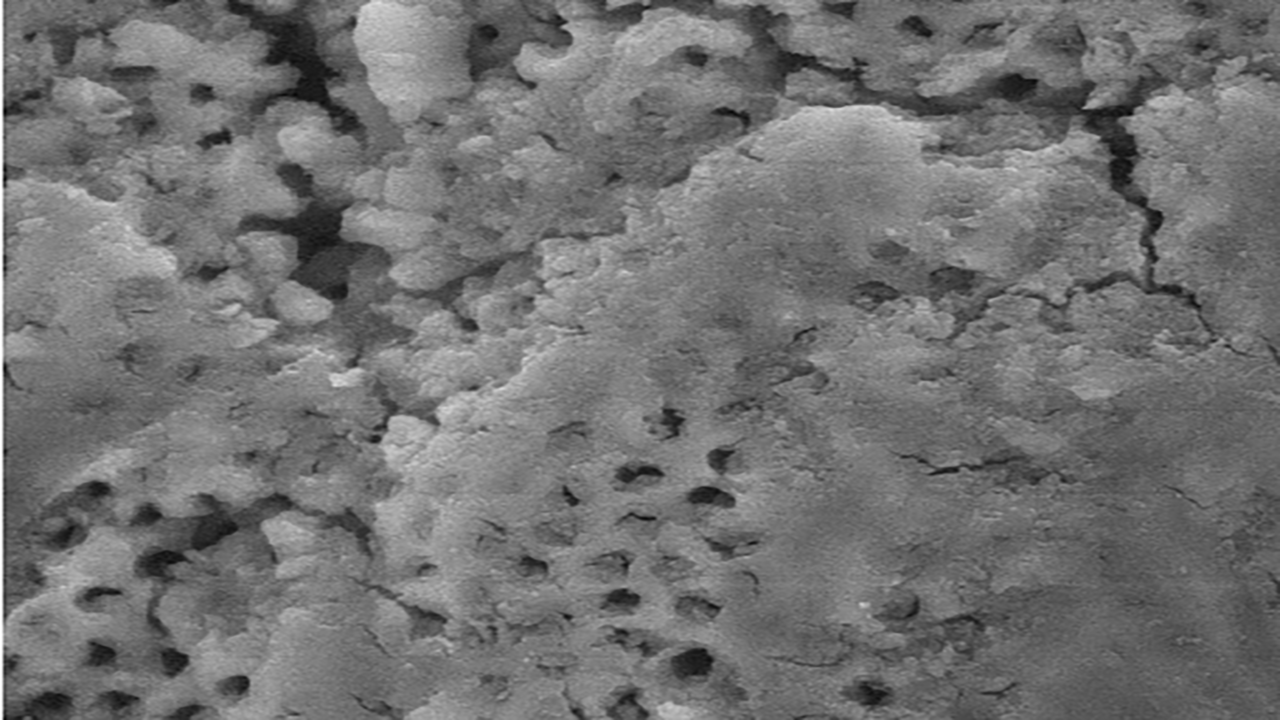
Whenever dentin is cut with either a hand instrument or a rotary instrument, the mineralised matrix shatters rather than being uniformly sheared or cleaved, producing considerable quantities of cutting debris. Much of the debris, made up of very small particles of mineralized collagen matrix, is spread over the surface of the dentin to form what has been called as smear layer.
According to Cohen, smear layer is defined as "an amorphous, relatively smooth layer of microcrystalline debris whose featureless surface cannot be seen with the naked eye".
Composition of Smear Layer
Organic portion contains - coagulated proteins, necrotic and non necrotic pulpal tissues, odontoblastic process, saliva, blood cells, micro-organisms and inorganic constituents are - Minerals from dentinal tubules.
Clinically produced smear layer have an average depth of 1 to 5 micrometer. Brainstorm described an inner and outer layer of smear layer in relation to dentin. The inner layer consisted of material which had been forced into the ends of dentinal tubules forming a smear plug. This sealed the tubule ends and decreases the permeability. The outer layer was an amorphous layer of 2.5 micrometer thickness lying on the actual surface covering the tubules and intertubular dentin.
Formation of Smear Layer
The normal structure of dentin comprises mineralized intertubular and peritubular dentin and dentinal tubules containing odontoblastic processes or their remnants and tissue fluid, often referred to as dentinal fluid. If the surface of cut enamel and dentin is examined after preperation with hand instruments or burs, no structural details such as cut dentinal tubules or enamel prisms will be visible, even at highest magnification. All such details are obscured by a covering layer of cutting debris from mineralized tissues. The grinding debris consists of ground components of enamel and intertubular and peritubular matrix, including any contents of the dentinal tubules, mixed with water, dentinal fluid and often saliva. This layer is less than 2 micrometer thick and termed as smear layer
Advantages and Disadvantages of Smear Layer
The smear layer is not a stable structure and it must be removed in order to obtain optimal chemical and mechanical bonding between restorative materials and tooth structures. This demineralization will allow resin to infiltrate the tubules and their branches, as well as the collagen meshbof the intertubular matrix and the collagen in the walls of the tubules exposed by the acid.
The presence of smear layer can be beneficial by physically reducing the flow through dentin and thus decreasing permeability. This reduced flow of dentinal fluid may have a protective effect on the pulpal tissues. This smear layer may also impede the entry of bacteria into the cut dentinal tubules. An alternative to removal of the smear layer for bonding to mineralized dental tissues is to incorporate it as an integral part of the adhesive system. Materials for such binding technique have been marketed.
Article by Dr. Siri P. B.


Hailey - 1 year ago