• Dentist Channel Online Dentist Channel Online
  • 23 April, 2022

Radiographic Assessment of Periodontium

The main features to be looked out are for

  • Crown/ Root Ratio 
  • Open contacts 
  • Root length and morphology,  
  • Local irritating factors 
  • Amount of bone present 
  • Condition of the alveolar crests 
  • Bone loss in the furcation areas 
  • Width of the periodontal ligament space 

Normal Anatomy 

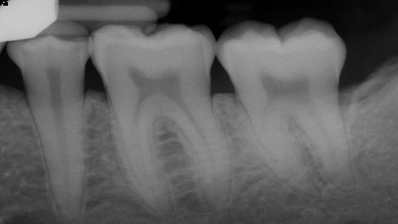
A thin layer of opaque cortical bone often covers the alveolar crest. The height of the crest lies at a level approximately 0.5 to 2.0 mm below the level of the CEJs of adjacent teeth. Between posterior teeth the alveolar crest also is parallel to a line connecting adjacent CEJs. Between anterior teeth the alveolar crest usually is pointed and may have a well-defined cortex. 

Between the anterior teeth, the normal alveolar crest is pointed and well corticated, coming to within 0.5 to 2.0 mm of the adjacent CEJs. The normal alveolar crest lies 0.5 to 2.0 mm below the adjacent CEJ and forms a sharp angle with the lamina dura of the adjacent tooth. 

The periodontal ligament (PDL) space is often slightly wider around the cervical portion of the tooth root, especially in adolescents with erupting teeth. In this situation, if the lamina dura still forms a sharp, well-defined angle with the alveolar crest, the condition is a variant of normal and is not an indication of disease. The thickness of alveolar crests varies and may be very thin coronally. This may appear radiographically as an increase in radiolucency toward the crest.  

The PDL varies in width from patient to patient, from tooth to tooth in the individual, and even from location to location around one tooth. Usually it is thinner in the middle of the root and slightly wider near the alveolar crest and root apex, suggesting that the fulcrum of physiologic movement is in the region where the PDL is thinnest. The shape of the tooth creates the appearance of a double PDL space. When the x-ray beam is directed so that two convexities of a root surface appear on a film, the double PDL space is seen. 

General Radiographic Features of Periodontal Disease 

  • Changes in the morphology of the supporting alveolar bone 
  • Loss of the interproximal crestal bone and bone overlapping the buccal or lingual aspects of the tooth roots
  • Changes to the internal density and trabecular pattern 
  • Changes to the internal aspect of the alveolar bone reflect either a reduction or an increase in bone structure or a mixture of both
  • Changes In Morphology Of The Alveolar Bone Appear as areas of localized erosion

 Article by Dr. Siri P. B.

Categories:
Tags :
Comments :
Hailey Avatar
Hailey - 1 year ago
My name is Hailey Garcia and I am from New Jersey. My herpes virus turned to war after 2 years of living with it. I have tried different medical procedures to cure my herpes but to no avail. Most people think herpes is only a minor skin irritation of which herpes has long term effects on health and passes through the bloodstream and can be easily contracted through sexual intercourse. I knew I had herpes from the first day I started feeling itchy in my pubic area and the pain was very unbearable. I couldn't stand it anymore. After 2 years of trying other means to get rid of it, I had to contact Doctor Odunga to help me with a permanent cure. I saw his email and whats-app number from a testimony I read online from a lady who was also helped by him in curing infertility problems, I had faith and contacted him. He assured me of his work and I ordered his herbal medicine. Within 5 days, I didn't feel any pain anymore and within 2 weeks, my skin was all cleared and smooth. I am very grateful to you sir and I write this testimony as others have done to bring those having faith to you sir. If you have herpes or other similar disease and you want it cured, kindly contact Doctor Odunga, Whats-App (wa.me/+2348167159012) OR Email [email protected]

Authentication required

You must log in to post a comment.

Log in

Upcoming webinars:

  • 1 April,2026
Introduction to Geriatric Dentistry & Geriatric Oral Healthcare

Introduction to geriatric dentistry focusing on oral healthcare for elderly patients.

  • 3 April,2026
Magnification in Dentistry: Entering the World of Precision

A webinar exploring how magnification tools like loupes and microscopes enhance precision, visualization, and clinical outcomes in modern dentistry.

  • 5 April,2026
Mastering Abutment Selection: The Key to Implant Success

A focused session on selecting the right implant abutment to enhance prosthetic fit, function, and long-term implant success.

  • 5 April,2026
Healing The Wounds –Sutures & Suturing

A clinical session on sutures and suturing techniques for effective wound closure and better healing in dental procedures.

  • 8 April,2026
Pink of oral health for the silver age

A concise webinar on oral healthcare and management strategies for the elderly population.

  • 14 April,2026
Dental Sleep Medicine: Next Blue Ocean in Dentistry

A webinar highlighting Dental Sleep Medicine and the dentist’s role in identifying and managing sleep-related breathing disorders like sleep apnea.

  • 15 April,2026
Breaking Silence: Social Health and Aging

A thought-provoking webinar addressing social health challenges, cultural taboos, and aging-related issues to improve holistic well-being in older populations.

  • 16 April,2026
Guided Endodontics for Calcified Canals – Predictability Redefined

A practical webinar on using guided endodontic techniques to improve predictability and success in managing calcified root canals.

  • 18 April,2026
Data to Decisions: Understanding Disease Measurement in Epidemiology

A webinar on understanding epidemiological disease measurement to interpret health data and support evidence-based decisions.