
 Dentist Channel Online
Crestal Bone Preservation
Authors – Marincola M.* Coelho PG** Morgan V.* Cicconetti A.**
* Ass. Professor, Department of Implant Dentistry, University Cartagena, Cartagena,
** Ass. Professor, Department of Biomaterials and Biomimetics, NYU.
* Clinical Director, Implant Dentistry Centre, Boston.
**Ass. Professor, Department of Oral Surgery, La Sapienza University, Rome.
Abstract
It is a general consideration to maintain bone around the dental implant. This is very necessary for the long-term success of the implant. In earlier times osseointegration was thought to an element of success for implant but it does not necessarily indicate that this bone material interface will keep its integrity throughout the patient life. There can be so many contributing factors for the bone loss. So, this article deals with all the factor related to crestal bone loss.
Introduction
It is general consensus that maintenance of bone around dental implants is one of the most important features in long term treatment success achievement, and that progressive bone loss drastically decreases the survivability of dental implants under occlusal loading.3,5 Although the achievement of Osseointegration after implantation is important in obtaining treatment success, it does not necessarily indicate that this bone biomaterial interface will keep its integrity throughout the patient’s life, since a large number of factors play a role in the kinetics of mineralized tissue. Of particular interest to the private practitioner is the Crestal bone loss after implantation occurring during the first year after implant placement. Once this loss will drastically affect the biomechanical anchorage of the prosthetic restoration and possibly jeopardize the proposed treatment. this loss may be attributed to several factors including,
a. excessive occlusal forces
b. trauma during the surgical procedure
c. inflammation/ infection
d. implant exposure during soft tissue healing
e. implant abutment gap present in the great majority of implant systems commercially available
f. early loading of a not biomechanically competent bone biomaterial interface
g. implant bulk device design, particularly the crest module profile
Among the potential causes described above, many can be avoided by the clinician through proper treatment planning and patient management, while others can be evaluated/avoided through engineering concepts. It is important to note that in most cases there is not a single factor but the synergy of various causes mediating the progressive mineralized tissue loss around dental implants. It is evident that full control of all the variable playing a role on bone loss around implants after implantation, especially around the implant crest module during the first year of implantation, is beyond any clinician’s domain, once there is contribution from the biological (patient), human technical (clinician) and engineering (implant designs and connectors) aspects to this complex problem.
Among the contributing factors that are in control of the clinician is a proper treatment planning to enable the right number and positioning of implants is a major issue. Once the proper number and position of implants is achieved, it facilitates proper restoration and occlusal adjustment thus diminishing to a great extent the human technical contribution to Crestal bone loss.
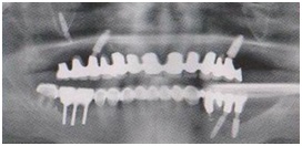
Proper treatment planning replacing the lost teeth with single crowns on short implants. Note the crest preservation over the intra-osseous Bicon Short Implants. (Fig 1)

Missing criteria for proper treatment planning. Poor implant placement with evidence of crestal bone loss. (Fig 2)
Another controlling factor is the engineering aspect of the implant system. The factors to be considered are the implant system. The factors to be considered are the implant crest module and the implant abutment connections. Although there is some evidence that both design considerations play a significant role on the Crestal bone loss around implants, quantification of these processes have not been experimentally shown to date due to multifactorial nature of the subject. The theories described in this series of articles rationalize Crestal bone loss related to crest module design and implant connections (particularly the one linking the implant to its respective abutment). These theories are in qualitative agreement with clinical observations for different implant designs.
The implant crest modules biomechanics:
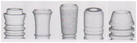
There are currently three different basic designs of implant crest modules available in commercial scale. These different geometries are shown in fig 3.
The three different crest module designs from left to right: “vase shaped cylindrical”, and “rocket shaped”. (Fig 3)

Schematic representation of commercially available implant systems and their respective crest modules are presented in figure 4. Throughout this article, the crest module in which the sides diverge to occlusal will be called “vase shaped” (VS), the one in which sides do not diverge or converge (parallel) “cylindrical” (C), and the one in which sides converge to occlusal will be called “rocket shaped” (RS). The qualitative static mathematical analysis regarding these three different crest module designs have been previously demonstrated during the late 80’s and is mentioned in several implant dentistry textbooks.
Commercially available implants with different crest module designs. From Left to Right: Noble BioCare (VS), ITI (VS), Astra Tech (VS), Ankylos (RS), and Bicon (RS).
The most desirable way to approach this type of problem is through mechanical and mathematical formulations with the aid of computer software (Finite Element Analysis), but qualitative understanding of the crest module role can be easily achieved through simple arguments on single tooth implant restorations as follows:
1. The forces that an implant is subjected during function are complex in nature due to the oblique planes comprising a crown, which make these forces oblique in nature thus resulting in vertical and horizontal force components. These vertical and horizontal force components will cause moments (force multiplied by the distance) in most instances, which may increase significantly the load to which the implant is subjected. Unless the load is vertical and perfectly aligned with the implant long axis, a horizontal force component acting on the implant will always exist.
2. Consider the schematic drawing of a vase shaped, a cylindrical and a rocket shaped implant in a bone domain as shown in Fig 5.
Implants on a bone domain of the same size. One should note the amount of bone present on the topmost part of the implant (presented by red arrows). The amount of bone around the crestal module is of fundamental importance on occlusal forces distribution. (Fig 5)

In these drawings, one should first note that the width of the bone domain is the same for all implant types and that these implants are inside this domain to their whole extent (the crest module is totally submerged in bone). It is also important that the implant diameters are the same (as if there were to be used to restore the same region).
It can be observed that in the cervical region of the crest module the bone amount around the module (red arrows) is smaller for the vase shaped implant than for the other two types, which leaves the vase shaped implant with a smaller amount of bone for force dissipation, showing that the bone around this crest module design is more likely to be overloaded and lost due to prosthetic occlusal function than the other two.6 This condition would be clinically accentuated in knife edged ridges, where a lesser amount of bone would be present around the implant’s crest module. This theory is in qualitative agreement with clinical findings where vase shaped implants present a slow but progressive bone loss after some implantation time in vivo and rocket shaped (biomechanically more favourable) implants present none or very little bone loss as time elapses in vivo (Fig 6-8).

Cone shaped crestal bone loss around a vase shaped(left) and a cylinder(right) implant neck after a period of loading. (Fig 6 & 7)
Note the crest preservation around the rocket shaped implant neck. The sloping shoulder guarantees a platform switching at implant level with bone growth over the neck. (Fig 8)
In spite of the higher amount of bone around the cervical part of the crest module of the cylindrical implant compared to the vase shaped implant, it has been shown by mathematical models which were in agreement with clinical observations have shown that there is an extensive progressive bone loss around implants presenting this geometry. This is likely due to the high interfacial shear stresses that these implants are subjected under vertical loading. For the other two crest module geometries, this progressive bone loss does not happen to the same extent and can be explained through simple mathematical calculations where the load vector (resultant) is broken in to components that are dependent on the crest module angulations, and interfacial shear stresses are attenuated when compared to cylindrical shaped crest modules. A simple representation of the reaction forces resulting from vertical load on a vase shaped implant is shown in fig 9 (vector magnitudes are not representative of their actual magnitude).
Decomposition of the reaction (dependent on the module geometry) resulting from a vertical load F applied on the implant. (Fig 9)

Further aggravation of the problem takes place as progressive bone loss occurs around the implants regardless of crest module design. As bone is lost (due to unfavourable biomechanical condition) from the upper part of the crest module downwards, implant anchorage is lost and there is an increase in load bearing of the remaining bone around the module due to an increase in the moment value (the moment increases proportionally to bone loss). This finding has been the subject of various laboratory and clinical research protocols, especially around vase shaped implant where theoretically this bone loss would evolve until catastrophic failure occurred, which is not the case. Interestingly, this bone loss usually stops at the first thread region and in most instances does not represent implant failure. In fact, these implants remain in place for long periods of time in function without any complications throughout its life time.8 This sudden stop at the first thread might be related to bacteriological contamination that may occur due to the presence of a gap on the implant abutment connection.4 Also this phenomenon has been taken in to account by several clinicians around the world, who have changed their surgical and restorative protocol to circumvent the drawbacks of such bone loss in order to achieve better results, especially in aesthetically compromised regions where the bone loss around the implant crest module makes handling of soft tissues difficult. It has been also reported by clinicians that the micro threads present on the crest module of an implant have significantly reduced bone loss (fig 10 and 11).
Comparison between a vase shaped (left) and a rocket shaped (right) implant design at crest level. (Fig 10 & 11)

Conclusion
In summary, it is widely accepted that the bone loss around implant’s crest module is multidisciplinary in nature and that from an engineering perspective these are related to device design (crest module design and implant connections). From a purely mechanical standpoint, if same diameter implants with the three-crest module designs available are to be placed in given region, the rocket shaped crest module implant will be less likely to lose bone around its crest module, which will theoretically help dissipating the functional loads.
It is paramount to remember that a long-term preservation of the Crestal bone makes the use of short implants predictable and encourage the clinician to use short implant in all kind of bone dimensions and bone quality. The rocket shaped module of a sloping shoulder can be considered as the ideal implant design for a homogeneous occlusal force distribution around the implant neck/Crestal bone.
In the past, it was believed that dental implants needed to be at least 10 mm in length to assure successful functioning of Osseo integrated implants however, recent studies show that short <10 mm dental implants can perform well. Particularly, the plateau or fin design of dental implants with a bacterially sealed 1.5-degree locking taper connection has provided for successfully functioning dental implants as short as 5mm in length. Additionally, they have shown that short unsplinted dental implants had less Crestal bone loss than longer splinted dental implants.
References
1. Bidez MW, Misch CE: Force transfer in implant dentistry: basic concepts and principles. J Oral Implant; 18:264-274, 1992.
2. Bozkaya D,Muftu S,Muftu A. Evaluation of load transfer characteristics of five different implants in compact bone at different load levels by finite elements analysis. J Prosthetic Dent 2004; 92:523– 530.
3. Horowitz, R., Current Implant Designs to Maintain Crestal Bone and Gingiva, Functional Esthetics & Restorative Dentistry: Series 1, Number 2, Dental Implants, p. 88-90, 200.
4. King GN, Hermann JS, School field JD, Buser D, Cochran DL. Influence of the size of the micro gap on crestal bone levels in non-submerged dental implants: a radiographic study in the canine mandible. J Periodontal 2002; 73:1111-1117.
5. Kitamura E,Stegaroiu R,Nomura S,Miyakawa O. Influence of marginal bone resorption on stress around an implant-a three-dimensional finite element analysis. J Oral Rehab 2005; 32:279– 286.
6. Lemons, J.E., Biomaterials, Biomechanics, Tissue Healing, and Immediate-Function Dental
Implants, Journal of Oral Implantology, Vol XXX No. 5 2004.
7. Leonard, G., Coelho, P., Polyzois, I., Stassen, L., Claffey, N., A study of the bone healing kinetics of plateau versus screw root design titanium dental implants, Clinical Oral Implants Research, 2009, 20 (3), 232-239.
8. Merickse-Stern, R., Aerni, D., Geering, A. H. and Buser, D. (2001), Long-term evaluation of no submerged hollow cylinder implants. Clinical Oral Implants Research, 12: 252–259.
9. Petrie CS, Williams JL. (2002): Shape optimization of dental implant designs under oblique loading using the p-version finite element method. J Mechanics in Medicine and Biology; 2(3-4): 339-45.
10. Shi Bin,Wu Leping,Wei Bin,Lin Gengbin,Huang Li,Qiu Yu Clinical Research of Straumann Implant System in Oral Prosthesis[J];Journal of Fujian Medical University;2007-06
11. Tada S,Stegaroiu R,Kitamura E,Miyakawa O,Kusakari H. Influence of implant design and bone quality on stress/strain distribution in bone around implants: a 3-dimensional finite element analysis. Int J Oral Maxillofacial Implants 2003;18:357–368.
12. Tawil G, Aboujaoude N, Younan R. Influence of prosthetic parameters on the survival and complication rates of short implants. Int J Oral Maxillofacial Implants 2006;21(2):275-282.
13. Venuleo, C., Chuang, S.K., Weed, M., Dibart, S., Long term bone level stability on Short Implants: A radiographic follow up study, Indian Journal of Maxillofacial and Oral Surgery, September 2008, Vol. 7: No.3, p. 340-345.
Titanium Implants Versus Zirconia Implants
Zygomatic Implants – Noris Medicals


Hailey - 8 months ago